Case Report - (2024) Volume 7, Issue 1
Dysphagia Caused by Anterior Cervical Osteophytes: A Rare Entity!
2Department of Surgery, Faculty of Medicine, University of Peradeniya, India
3Teaching Hospital Peradeniya, India
Received Date: Dec 20, 2024 / Accepted Date: Jan 30, 2024 / Published Date: Mar 21, 2024
Abstract
When we go through the medical literature, anterior cervical osteophytes are generally considered to be asymptomatic. However, on multiple occasions, there have been reports of large anterior cervical osteophytes giving rise to dysphagia in elderly patients. In our article, we will be discussing a 79-year-old male patient who presented with dysphagia. We thoroughly evaluated this patient for a possible cause of his symptoms. He had to undergo multiple investigations, including an upper-gastrointestinal endoscopy, a lateral neck X-ray, and a contrast-enhanced computed tomography scan of the neck and chest. As well, we also arranged neurology and neurosurgical referrals. Ultimately, we could confidently conclude that he was having dysphagia due to anterior cervical osteophytes. Other than patient evaluation, we have also briefly discussed in this article the treatment modalities and how to choose the best treatment modality for a patient with symptomatic anterior cervical osteophytes.
Categories: General Surgery, Orthopedics, Neurosurgery
Keywords
Diffuse Idiopathic Skeletal Hyperostosis (DISH), Anterior Cervical Osteophytes (ACOS), Dysphagia, Osteophytectomy
Introduction
Although anterior cervical osteophytes (ACOs) are not an uncommon entity in the elderly, they are rarely identified as an etiology for dysphagia in the surgical literature [1]. Diffuse idiopathic skeletal hyperostosis (DISH) and cervical spondylosis are two pathological conditions that give rise to anterior cervical osteophytes [1, 2], but most of the anterior cervical osteophytes occur in the degenerative diseases of the spine, where they are formed by reactive ossification of the anterior longitudinal ligaments [3]. Anterior cervical osteophytes should be considered as a cause of dysphagia only after excluding other pathological conditions of the esophagus, such as tumors, gastric ulcers, strictures, achalasia, and neuromuscular diseases [3]. This case report is regarding a 79-year-old male patient with dysphagia due to osteophytes of the cervical spine.
Case Presentation
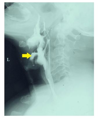
A 73-year-old male patient presented with a history of suddenonset dysphagia for one month after an episode of sore throat. Dysphagia was there for both solids and liquids equally. He complained of a sticking sensation at the level of the neck region. There was occasional nasal regurgitation of food. He had lost about 2 kilograms of weight during the last month due to poor food intake. Dysphagia is said to occur a few seconds after swallowing a food bolus. He did not complain of any loss of appetite, vomiting of blood, episodes of per-rectal bleeding, or other sinister symptoms. He has not had any family history of similar symptoms or a family history of malignancies. On the general examination, he had no significant findings other than a low body mass index of 17.85 kg/m2. His cardiovascular, respiratory, and nervous systems and abdominal examination findings were unremarkable. We were unable to find any palpable cervical lymph nodes during the head and neck examination. He initially underwent an upper-gastrointestinal endoscopy. We were unable to enter the esophagus due to a soft tissue prominence in the posterior wall of the pharynx. No other gross abnormalities were detected during the upper gastrointestinal endoscopy, and the vocal cords were normal. An x-ray of the lateral neck was performed as the preliminary investigation. Sharp cervical osteophytes at C4 and C5 were detected squeezing the pharynx (Figure 1).

Figure 1: X-ray lateral neck Sharp cervical osteophytes at the levels of C4 and C5 are shown by the yellow arrow
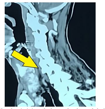
A contrast-enhanced computed tomography (CECT) scan of the neck and chest was performed (Figure 2). It revealed ossification of the anterior longitudinal ligament from C2 to upper thoracic vertebral levels, impinging on the pharynx and esophagus markedly at the levels of C4 and C5. In conclusion, the appearance was in favor of dysphagia secondary to diffuse idiopathic skeletal hyperostosis (DISH). As well, the CECT study also excluded other possible extraluminal and intraluminal causes, such as neoplasms.

 Figure 2: CECT neck and chest Osteophytes impinging on the pharynx and esophagus at the levels of C4 and C5 (shown by the yellow arrow).
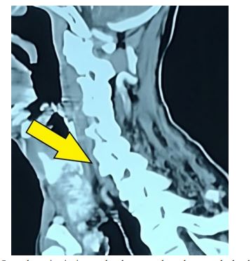
An upper gastrointestinal contrast study was also performed (Figure 3). It revealed an irregularity of the posterior pharynx caused by degenerative changes in the cervical spine. This study also revealed contrast passage into the vestibule during swallowing, likely due to reduced motility, particularly that of the epiglottis.

Figure 3: Upper GI Contrast Study (Contrast swallow) Contrast passage into the vestibule during swallowing is show by the yellow arrow.
Neurology and neurosurgical referrals were done to exclude the possibility of a neurological disorder. Reporting said there are no features that suggest a neurological cause for dysphagia. They concluded that mechanical compression with DISH in the cervical spine was the cause of dysphagia.
Results and Discussions
The prevalence of anterior cervical osteophytes in the elderly ranges from 20%-30%, mostly seen in males [1]. The majority are asymptomatic, and large osteophytes causing dysphagia are rare. Cervical spine degeneration, cervical spondylosis, or DISH may all result in large anterior cervical osteophytes [2].
Anterior cervical osteophytes-related dysphagia has been explained by a variety of theories, such as esophageal obstruction caused by mechanical compression, esophageal fibrosis, peripharyngitis and periesophagitis, cricopharyngeal spasms due to pressure on the esophagus, impaired motility of the epiglottis, and laryngeal cartilage distortion from osteophytes [2].
Since anterior cervical osteophytes do not commonly cause dysphagia, a diagnosis of this kind should only be established after all other possible common causes of dysphagia have been ruled out. Before the diagnosis of ACOs is made, all patients should have a routine diagnostic workup for dysphagia to rule out intrinsic causes such as diverticula, oesophageal stricture, motility problems, and neoplasms [3]. For most patients, the diagnosis is established by a thorough history, indirect laryngoscopy, and lateral cervical spine X-ray images [1]. Upper gastrointestinal endoscopy can be performed in patients with cervical osteophytes, but cautiously, considering the risk of esophageal perforation [4]. So, surgeons have to be aware of the possibility of esophageal perforation when performing upper gastrointestinal endoscopy on the elderly with dysphagia. A barium swallow or upper gastrointestinal contrast study (contrast swallow) may confirm the obstructive nature of the osteophytes. Excluding motility problems and gastro-esophageal reflux disease as potential causes of neck dysphagia may be aided by esophageal manometry and pH stimulation tests [5]. The diagnosis can be confirmed by a contrast- enhanced CT scan of the neck and thorax.
All individuals with dysphagia due to ACOs should be provided conservative therapy once a diagnosis has been made [3]. Diet modification (soft diet) along with anti-reflux or antiinflammatory drugs, muscle relaxants, and swallowing therapy has been shown to be beneficial in patients with ACOs with mild symptoms [6]. Patients who do not improve with conservative management are candidates for surgery [3].
These individuals may be treated using either anterolateral or posterolateral transcervical approaches to surgically excise the anterior cervical osteophytes (osteophytectomy). According to studies, both methods have provided equally effective dysphagia palliation without related morbidity. The anterolateral approach seems to be better for safeguarding the carotid sheath, despite the increased risk of injury to the recurrent laryngeal nerve. On the other hand, the posterolateral approach provides more access to prevertebral space, but it also raises the risk of sympathetic chain damage and necessitates more carotid sheath retraction. Depending on the attending surgeon's discretion, either exposure may be employed [7].
Nevertheless, following surgery, postoperative hematoma and laryngeal edema have been documented to cause a transient increase in dysphagia and hoarseness of voice [8]. Furthermore, following surgery, the condition will invariably recur [8]. So, in this case, to prevent unanticipated morbidity, the scheduling of surgery for the treatment of dysphagia should always be closely evaluated at an individual level [3].
Conclusions
Although uncommon, large anterior cervical osteophytes are a known cause of dysphagia, which is primarily observed in older adults. All patients should have a complete assessment before the diagnosis of anterior cervical osteophytes is established to rule out more prevalent and sinister causes of dysphagia. For any patient experiencing dysphagia as a result of anterior cervical osteophytes, conservative therapy should be provided. After carefully weighing the advantages over the disadvantages, patients who are not responsive to conservative treatment and those who have severe and worsening symptoms should be offered surgical removal of cervical osteophytes.
References
1. Rao, V., Shankar, R., Shivappa, N., & Nagaraj, T. M. (2014). Dysphagia caused by anterior cervical osteophyte: A rare entity revisited. International Journal of Head and Neck Surgery, 3(3), 168-171.
2. Giger, R., Dulguerov, P., & Payer, M. (2006). Anterior cervical osteophytes causing dysphagia and dyspnea: an uncommon entity revisited. Dysphagia, 21, 259-263.
3. Kuruwitaarachchi, K., Bandara, S. C., & Atthanayake, D. (2021). Progressive dysphagia secondary to multiple anterior cervical osteophytes: a case report. Journal of the Postgraduate Institute of Medicine, 8(2), 1-4.
4. Wright, R. A. (1980). Upper-esophageal perforation with a flexible endoscope secondary to cervical osteophytes. Digestive diseases and sciences, 25, 66-68.
5. Srinivas, P., & George, J. (1999). Cervical osteoarthropathy: an unusual cause of dysphagia. Age and ageing, 28(3), 321- 322.
6. Unlu, Z., Orguc, S., Eskiizmir, G., Aslan, A., & Tasci, S. (2009). The role of phonophoresis in dyshpagia due to cervical osteophytes. International journal of general medicine, 11-13.
7. Carrau, R. L., Cintron, F. R., & Astor, F. (1990). Transcervical approaches to the prevertebral space. Archives of Otolaryngology–Head & Neck Surgery, 116(9), 1070-1073.
8. Ruetten, S., Baraliakos, X., Godolias, G., & Komp, M. (2019). Surgical treatment of anterior cervical osteophytes causing dysphagia. Journal of Orthopaedic Surgery, 27(2), 2309499019837424.
Declarations
The patient gave full permission for the publication of this case report and the use of photographs of the relevant x-rays and the other investigations. Written consent is available with me as the corresponding author.
Copyright: © 2025 This is an open-access article distributed under the terms of the Creative Commons Attribution License, which permits unrestricted use, distribution, and reproduction in any medium, provided the original author and source are credited.


