Research Article - (2022) Volume 7, Issue 2
A better diagnose for canine pathology patients by other associated dental anomalies
Received Date: Jan 24, 2022 / Accepted Date: Jan 31, 2022 / Published Date: Feb 10, 2022
Abstract
Background: Canine pathology is one of the most difficult anomalies found in orthodontic patients, mostly when we must deal with an impacted canine. This is the reason why we, as general dental practitioners, pedodonts or orthodontists should be able to diagnose any canine pathology as soon as possible, so that the patient could follow interceptive treatment.
Methods: This is an observational study based on two retrospectives studies held in a private practice, Orto Office, in Bucharest, Romania. An amount of 400 complete orthodontic records were searched for impacted canine and ectopic canine.
Results: We found 54 patients responding to our criteria, 14 with impacted canine and 40 with ectopic canine. Most impacted canines in our study group were vertical 78.5%. The most frequent place for canine ectopy was found to be vestibular in 95% of the cases. In 64.3% of the impacted canine cases another dental anomaly was also diagnosed. For the canine ectopy group no dental anomaly was detected in 90% of the patients.
Conclusion: Our present findings are important especially for early diagnosing canine pathologies as the other dental anomalies may be observed earlier: the nanic lateral incisor or its anodontia. Moreover, we can prevent the canine impaction or ectopy by treating patients in their early ages around 8-10 years of age. These dental anomalies should be seen not only as risk indicators for early diagnosis but also as possible means of diagnose.
Keywords
Canine Pathology, Dental Anomalies, Canine Impaction, Canine Ectopy, Anodontia
Introduction
Any tooth that is unerupted more than one year after the normal period of its eruption is identified as ‘retained’ [1]. The imposibillity of the tooth to come out on to the dental arch, frequently due to either lack of space or the existence of an entity impeding its normal path of eruption, outcomes to dental impaction [2].
Canine pathology raises among the most difficult anomalies to treat in orthodontic patients, mostly when we must deal with an impacted canine. This is the reason why we, as dental general practitioners, pedodonts or orthodontists should be able to diagnose any canine pathology as soon as possible, so that the patient could follow interceptive treatment. In order for that to happen, we need to rise awareness among others of the importance of early dentition signs of lack of space, so that the later canine pathology could pe prevented or ameliorated. Of course, later on, we would at least diagnose as soon as growth is not over yet, so that the treatment would be shorter and have a better stability in retention; that is why we need to look for other signs that could lead us to the correct diagnostic.
Purpose
The aim of this study is to find enough data on dental anomalies associated with canine ectopy or impaction to have better chances to diagnose and treat these canine pathologies as soon as possible and even prevent them when chances are high. It is also highly significant to compare our outcomes to other surveys in other countries from all over the world. We aim to have a better diagnose for canine ectopy and canine impaction, by early diagnosing other dental anomalies like lateral incisor anodontia or nanic lateral incisor
Materials and Methods
This is an observational study based on two retrospectives studies held in a private practice, Orto Office, in Bucharest, Romania. An amount of 400 complete orthodontic records consisting of: three facial pictures, six dental pictures, orthopantomograms and teleradiographs were analyzed. The records included in this study were gathered on an eight-year period, between 2013 and 2021.
The inclusion criteria were any patient regardless of sex with at least one ectopic or impacted canine (upper or lower). We found 54 patients corresponding to our criteria, 14 with impacted canine and 40 with ectopic canine. All the impacted canine patients regarded as adequate for the present study, were carefully analyzed for the following data: age, sex, type of impaction (horizontal or vertical), skeletal class, occlusal transverse relation, vertical anterior relation, palate form, other associated dental anomalies. All these data were gathered in a table.
The ectopic canine group was also closely checked up for: age, sex, type of ectopy (buccal or palatal), skeletal class, occlusal transverse relation, vertical anterior relation, palate form, other dental anomalies associated. The data was also written down in one table.
Statistical managing of the information from the present research needed the following programs: the Microsoft Excel from Microsoft Office 2015, Google Docs and Google Drive.
All patients qualified for the present survey signed a consent form in which they approve to the use of their pictures, radiographs, and other examinations for scientific exploration.
Results
It has been found that 54 out of 400 patients had an ectopic or an impacted canine, 13.5%. The study didn’t take into consideration the complete transposition cases.
The age repartition was similar in both groups: around half of the patients were under 18 years: 57.1% for the impacted canine group and 50% for the ectopic canine group.
The sex distribution was surprisingly different: for the impacted canine group we found 57.1% men while for the ectopic canine group, the women had majority with a percentage of 77.5%.
Most impacted canines in our study group were vertical 78.5%. Only 21,5% of the cases were horizontal canine impaction (regarding the α angle). The inclusion measurement for whether horizontal or vertical impaction was in this case the α angle on the panoramic x-ray: α-angle is measured between the long axis of the impacted canine and the midline [3,4] as it is highlighted in figure 1 and figure 2.
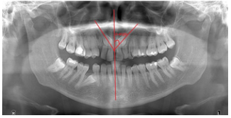
Figure 1: α-angle measurement.
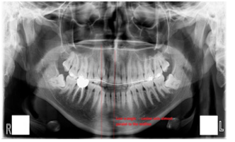
Figure 2: Low α-angle (almost 0).
The most frequent place for canine ectopy was found to be vestibular in 95% of the cases that we have investigated (figure 3).
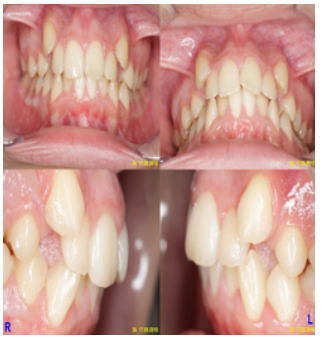
Figure 3: Vestibular canine ectopy
The skeletal features are very similar in both groups as it was expected, the canine pathology, whether it is ectopy or impaction it doesn’t affect the skeletal type and neither the vice versa isn’t happening. So, for the ectopy study group, 45% of the patients were class I, 35% were class II and only 20% were class III. Regarding the impacted canine group, half of the patients were class I Angle and 35.7% were class II, while 14.3% skeletal class III Angle. This outcome is in some way expected as most of the patients in our area are class I or II, the incidence of skeletal class III patients is quite low in Romania.
Further along our study we compared the two groups regarding dental occlusion: saggital, transverse and vertical, all the data was gathered in table 1.
The list of abbreviations used in the table are explained in below.
I-Angle class I
II-Angle class II
III-Angle class III
CB-Cross bite
ICB-Incomplete cross bite
N-Neutral occlusion
L-Lingualized occlusal relations
DB-Deep bite
ETE-Edge to edge occlusal relations
ACB-Anterior cross bite
Table 1
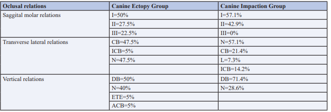
Further, we looked for other dental anomalies associated with canine ectopy or canine impaction to be able to draw a connection between a dental anomaly that is early diagnosed (like nanic lateral incisor) and canine impaction or canine ectopy-anomalies usually detected too late when they are difficult to treat. Table 2 was conceived to outline the link between dental anomalies like anodontia or nanic lateral incisor and canine ectopy and canine impaction. The results showed that in 64.3% of the impacted canine cases another dental anomaly was also diagnosed, whether it meant anodontia - figure 4, supernumerary teeth, ectopy or nanic lateral incisors.
Table 2

For the canine ectopy group no dental anomaly was detected in 90% of the patients.
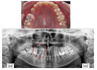
Figure 4: Both upper left premolar anodontia and impacted upper canine.
Discussion
In the recent past we have conducted similar studies to observe the two different canine pathologies and our conclusions were similar to this present study [6,7].
Our previous and present studies show that there is a connection between impacted canines and other dental anomalies and these facts concur with the work of Carvahlo et al [7], Paschos et al [8], Peck et al [9], Sacerdoti [10], Leifert [11], Langberg [12], Mesotten et al [13], Nagpal et al [14]. The reason for this incidence in the specialty literature about this relation is that the surplus of space in the maxilla could be a contributing factor for the canine misplacement and subsequently impaction as it allows enough space for the canine to depart its normal direction of eruption. Alternatively, the lack of eruption guidance from the lateral incisor, enables a new direction for the canine to take to the palatine side. A different likely explanation is the potential genetic connection between impacted canine and tooth size decrease. The theory is that dental anomalies of number, tooth size reduction and impacted canines are three of the substitutes in a genetically controlled dental conditions appearing on a regular basis together. It is probable that the gene (or genes) to blame for the management of teeth eruption and afterwards for the palatal displaced canines are related to the gene (or genes) that establish hypodontia or incisor agenesis. Our present findings are very important especially for early diagnosing canine pathologies as the other dental anomalies may be observed earlier, for example the nanic lateral incisor or its anodontia. Moreover, we can prevent the canine impaction or ectopy by treating patients in their early ages around 8-10 years of age. These other anomalies should be seen not only as risk indicators for early diagnosis as Herrera-Atoche [15] concludes but also as possible diagnostic means.
Rqafeeq [16] et al believes that orthodontists should consider any type of delay in canine eruption thoroughly and seriously especially when other dental anomalies as dental agenesis. It is imperative that once a dental anomaly is detected in one young patient, he should be monitored through the permanent teeth eruption to prevent canine impaction or ectopy. The patient should be advised to see an orthodontist as early as possible to be able to prevent these difficult pathologies.
Treating an impacted canine is among most challengeable orthodontic treatments according to Kokich [17], so any early sign should be taken into consideration and any methods of preventing this affection should be prioritized by general dentists and orthodontists. Therefore, there is a great need of rising awareness whenever we see a 8 or 9 year old with nanic lateral incisor or we suspect lateral incisor anodontia, as there are 35,7% chances to suffer from impacted canine also.
Conclusion
For a better and a faster early diagnose of impacted canines or canine ectopy, we need a screening for children 8-9 years of age and closely monitor the patients with lateral incisor anodontia or nanism and refer them to an orthodontist.
On the other hand, orthodontists should consider any type of delay in canine eruption thoroughly and seriously especially when also suffering from other dental anomalies as dental agenesis. It is imperative that once a dental anomaly is detected in one young patient, he should be observed through the permanent teeth eruption to prevent canine impaction or ectopy.
References
1. Torres-Lagares D, Flores-Ruiz R, Infante-Cossio P, GarciaCalderon M, Gutierres-Perez JL (2006) Transmigration of impacted lower canine. Case report and review of literature. Medicina Oral, Patologia Oral y Cirugia Bucal 11:171-174.
2. Daskalogiannakis J, Multilingual glossary of orthodontic terms, 2000 Berlin Quintessence Publishing Co, Inc.pg. 142.
3. Belma I√Ö?√Ą¬Īk Aslan and Neslihan Üçu√Ć?ncu√Ć? (March 11th 2015). Clinical Consideration and Management of Impacted Maxillary Canine Teeth, Emerging Trends in Oral Health Sciences and Dentistry, Mandeep Singh Virdi, IntechOpen, DOI: 10.5772/59324. Available from: https://www. intechopen.com/chapters/47825
4. Turk MH, Katzenell J (1970) Panoramic Localization. Oral Surgery, Oral Medicine and Oral Pathology 29(2) 212-215.
5. Moro√Ö?an H (2021) Perspectives on Prevalence of Canine Ectopy in Romanian Orthodontic Patients. Int J Medical Dentistry 25(2).
6. Moro√Ö?an H (2022) Canine Impaction and Other Dental Anomalies – What is the Connection? Journal of Dentistry and Oral Biology7(1).
7. Carvalho, Anísio Bueno de, Motta, Rogério Heladio Lopes and Carvalho, Eliane Maria Duarte de (2012) Relation between agenesis and shape anomaly of maxillary lateral incisors and canine impaction. Dental Press Journal of Orthodontics 17:83-88.
8. Paschos E, Huth KC, Fässler H, Rudzki-Janson I (2005) Investigation of maxillary tooth sizes in patients with palatal canine displacement. J Orofac Orthop 66(4):288-298.
9. Peck S, Peck L, Kataja M (2002) Concomitant occurrence of canine malposition and tooth agenesis: evidence of orofacial genetic fields. Am J Orthod Dentofacial Orthop. 2002;122(6):657-60.
10. Sacerdoti R, Baccetti T (2004) Dentoskeletal features associated with unilateral or bilateral palatal displacement of maxillary canines. Angle Orthod 74(6):725-732.
11. Leifert S, Jonas IE (2003) Dental anomalies as a microsymptom of palatal canine displacement. J Orofac Orthop 64(2):108- 120.
12. Langberg BJ, Peck S (2000) Tooth-size reduction associated with occurrence of palatal displacement of canines. Angle Orthod 70(2):126-128.
13. Mesotten K, Naert I, van Steenberghe D, Willems G (2005) Bilaterally impacted maxillary canines and multiple missing teeth: a challenging adult case. Orthod Craniofac Res 8(1):29- 40.
14. Nagpal A, Pai KM, Sharma G (2009) Palatal and labially impacted maxillary canineassociated dental anomalies: a comparative study. J Contemp Dent Pract 10(4):67-74.
15. Herrera-Atoche JR, Agüayo-de-Pau M, Escoffié-Ramírez M, Aguilar-Ayala FJ, Carrillo-Ávila BA, Rejón-Peraza ME (2017) Impacted maxillary canine prevalence and its association with other dental anomalies in a Mexican population. Int J Dent 2017:7326061.
16. Rafeeq RA, AL-mashhadany SM, Hussein HM, AL-sudani rj, ali mqm (2021) The relation between maxillary buccal canine impaction and dental anomalies-A retrospective study. International Journal of Pharmaceutical Research 13(1).
17. Kokich VG, Mathews DP (2014) “Orthodontic and Surgical Management of Impacted Teeth”, Quintessence Publishing 27-102,.
Copyright: © 2025 This is an open-access article distributed under the terms of the Creative Commons Attribution License, which permits unrestricted use, distribution, and reproduction in any medium, provided the original author and source are credited.


